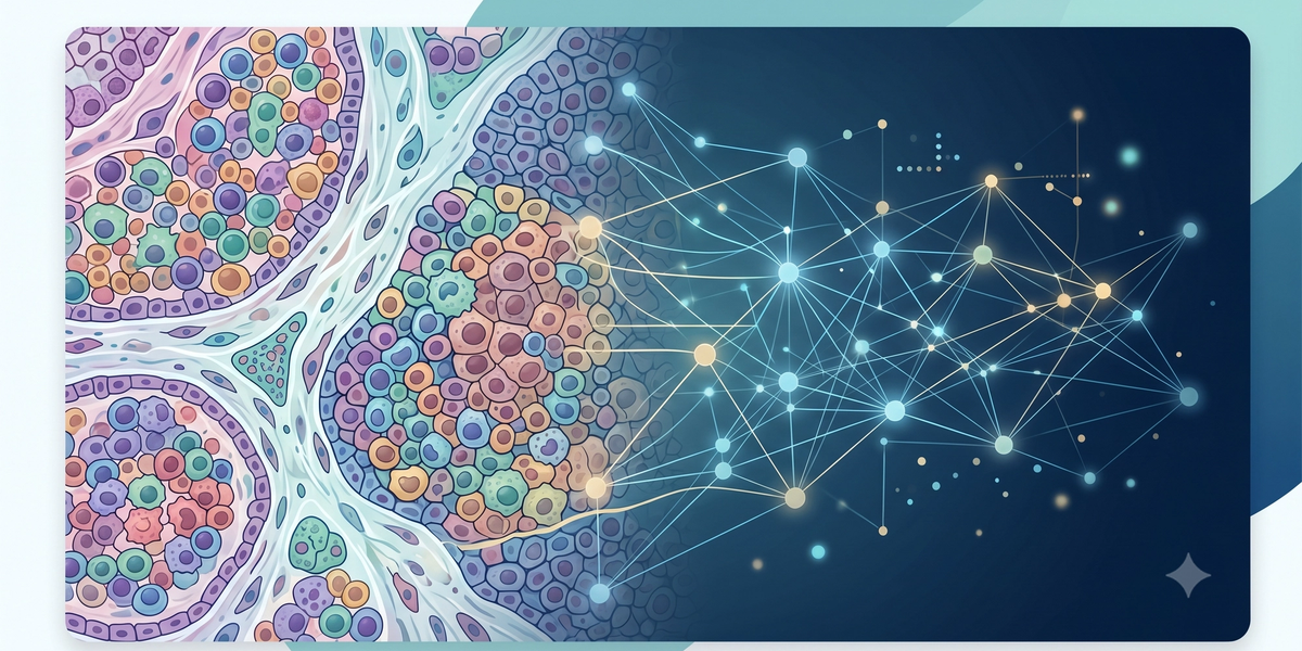
AI and Cancer Detection - Research Project
Discover how modern AI algorithms and spatial pattern detection are transforming oncology by analyzing the tumor microenvironment to identify, map, and understand cancer risk better than ever before.
Cancer research is no longer limited to studying the tumor alone. In colorectal cancer, the area surrounding the tumor — known as the tumor microenvironment — can strongly influence how the disease behaves. This environment includes immune cells, stromal tissue, blood vessels, and other surrounding structures that interact with cancer cells. Recent research highlights that colorectal cancer progression is shaped by the interaction between tumor cells and the tumor microenvironment.
This is where artificial intelligence can play an important role. Instead of only identifying whether a tissue region is cancerous, AI can help analyze how different tissue types are arranged around the tumor. For example, the distance between immune cells and tumor regions, the density of lymphocytes, and the amount of supportive stromal tissue may reveal patterns linked to disease severity.
The attached MachinedMind research proposal focuses exactly on this idea: using AI to classify tissue types from histopathology images, study spatial patterns between tumor regions, lymphocytes, and stroma, and build a simple mathematical risk-scoring framework.
Why Spatial Analysis Matters in Cancer Research
Two patients may have tumors that look similar under a microscope but still experience very different outcomes. One possible reason is that the surrounding tissue environment may be different. Research suggests that the location and behavior of immune cells within the tumor microenvironment can have prognostic value in colorectal cancer.
For a student research project, this opens a powerful question:
Can AI detect hidden spatial patterns in cancer tissue that may help explain patient risk?
By using machine learning on annotated histopathology images, students can explore features such as:
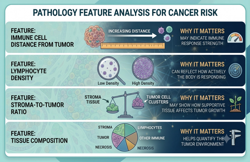
AI-enabled spatial analysis of whole-slide images has already been used in colorectal cancer research to characterize the tumor microenvironment more deeply.
What Students Can Build in This Project
A student working on this topic can build a strong interdisciplinary portfolio by combining biology, AI, mathematics, and medical data analysis.
The project can include:
- Tissue Classification Model
Use a pre-trained AI model to classify regions as tumor, lymphocytes, stroma, or other tissue types. - Spatial Feature Extraction
Measure distances, densities, and ratios between tissue regions. - Risk Score Framework
Create a simple weighted score using mathematical features such as immune-cell proximity, lymphocyte density, and stromal proportion. - Research Paper
Convert the findings into a structured research paper suitable for high school-level competitions or publication platforms.
This approach is especially useful because modern AI methods can support the discovery of spatial biomarkers from routine histopathology images.
Why This Is a Strong Research Topic for High School Students
This topic is advanced, but it can be made student-friendly with the right guidance. It does not require the student to conduct wet-lab experiments. Instead, the student can work with public datasets, Python, machine learning libraries, and Google Colab.
The project also teaches important research skills:
Literature review, medical image analysis, AI model interpretation, statistical reasoning, and scientific writing.
For students interested in medicine, biotechnology, computer science, or data science, this kind of project can become a strong portfolio piece because it shows the ability to apply AI to a meaningful real-world healthcare problem.
MachinedMind Angle
At MachinedMind, students can work on research projects that go beyond basic literature review. A project like this can be developed into a complete research experience: from understanding colorectal cancer biology to building an AI-based analysis pipeline, creating a risk-scoring model, and writing a structured research paper.
Final takeaway: AI can help students move from simply “studying cancer” to asking a deeper research question — can hidden tissue patterns around a tumor help us understand cancer risk better?
References
- Kennel, K. B., & Greten, F. R. (2025). The immune microenvironment of colorectal cancer. Nature Reviews Cancer, 25, 945–964. doi:10.1038/s41568-025-00872-1
- Lim, Y., Choi, S., Oh, H. J., et al. (2023). Artificial intelligence-powered spatial analysis of tumor-infiltrating lymphocytes for prediction of prognosis in resected colon cancer. npj Precision Oncology, 7, 124. doi:10.1038/s41698-023-00470-0
- Wu, X., Yan, H., Qiu, M., et al. (2023). Comprehensive characterization of tumor microenvironment in colorectal cancer via molecular analysis. eLife, 12, e86032. doi:10.7554/eLife.86032
- Tsai, P. C., Lee, T. H., Kuo, K. C., et al. (2023). Histopathology images predict multi-omics aberrations and prognoses in colorectal cancer patients. Nature Communications, 14, 2102. doi:10.1038/s41467-023-37179-4
- Idos, G. E., Kwok, J., Bonthala, N., et al. (2020). The prognostic implications of tumor infiltrating lymphocytes in colorectal cancer: A systematic review and meta-analysis. Scientific Reports, 10, 3360. doi:10.1038/s41598-020-60255-4

MachinedMind Team